Följ med på en resa långt, långt bort
Följ med på en resa ut genom Vintergatan. Vi reser runt bland galaxer och kvasarer, ända ut till Big Bang.
Liveföreställning i OpenSpace
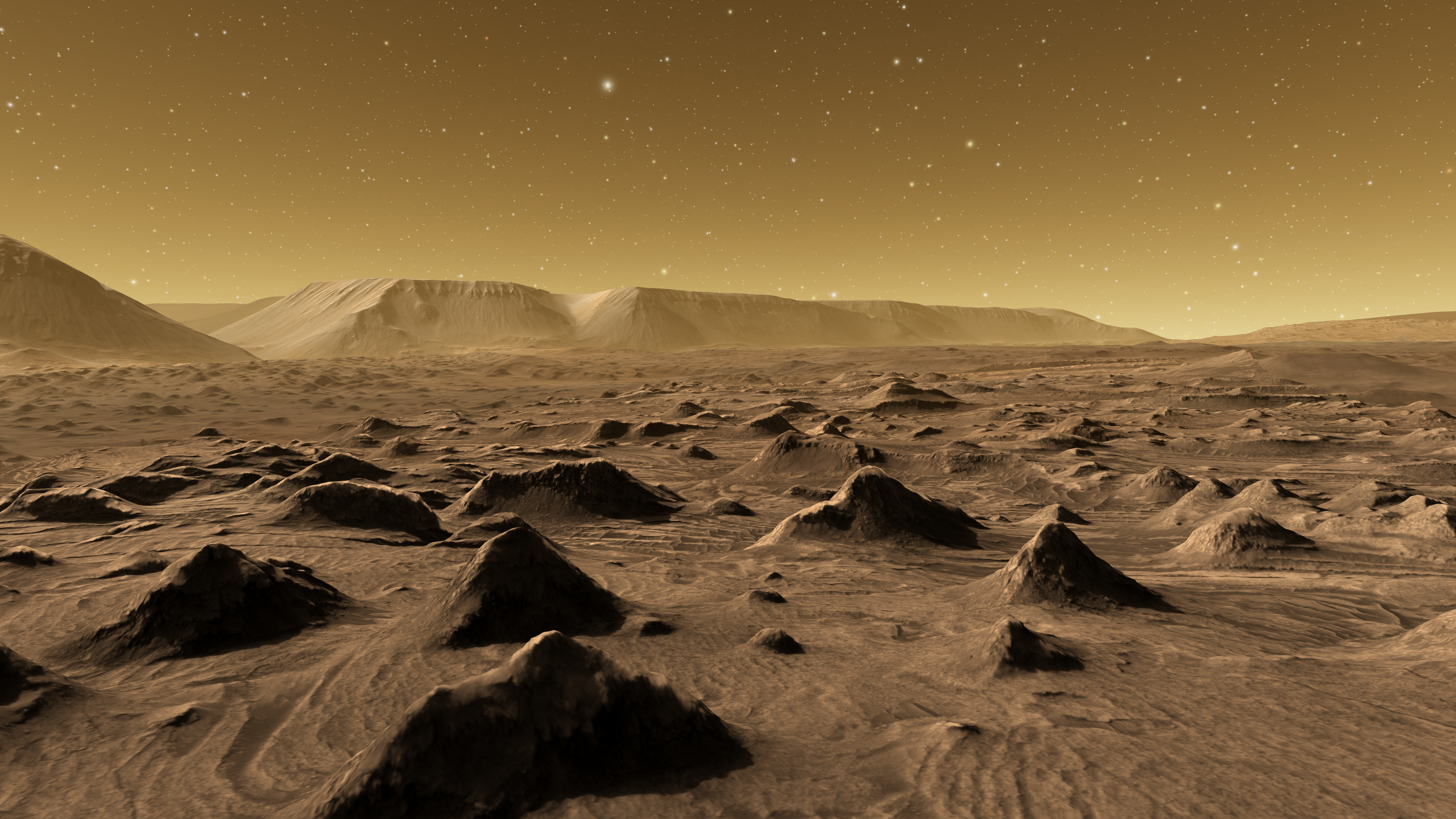
Under En kvart i rymden får du följa med på en spännande liveföreställning för hela familjen. Resan är ca 20 minuter lång och startar i vår VR-Arena där du möts av biografstolar och en häftig, platt storbildskärm i 2D. Resan fylls av sekvenser och bilder från astrovisualiseringsprogrammet OpenSpace.
OpenSpace är framtaget av Visualiseringscenters forskare i samarbete med NASA och American Museum of Natural History. Med hjälp av realtidsdata och vetenskapliga fakta om astronomi och rymden visualiserar programmet hela det kända universum.
Rekommenderad ålder
Den här föreställningen passar för hela familjen. Välkommen!
Information
Plats: VR-arenan, plan 1
Format: 2D, flatscreen
Tid: Ca 20 minuter
Pris: Gratis
Åldersgräns: Från 8 år
Bokning: Begränsat antal platser. Bokas i förväg via webbsida eller på plats.